
CSF hypotension
Findings:
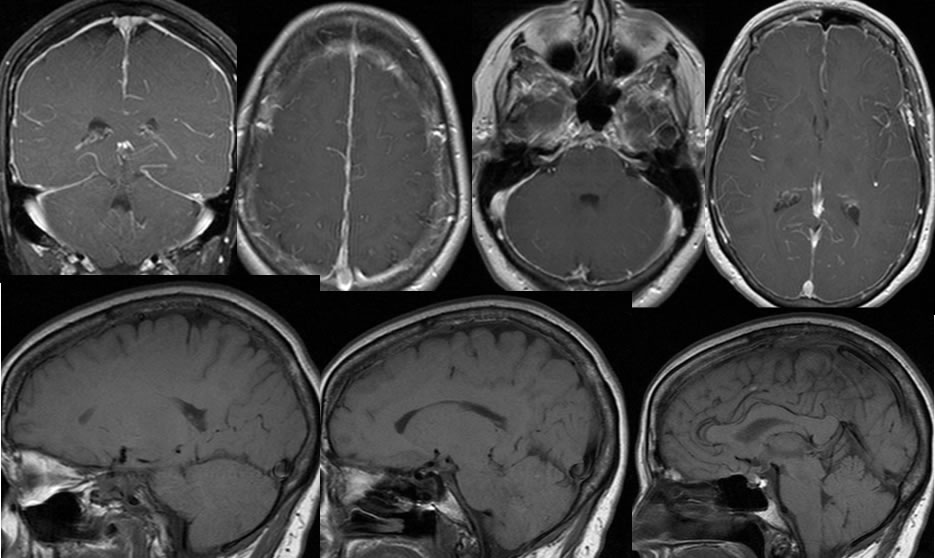
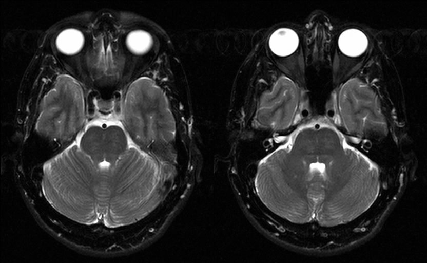
Diffuse uniform dural enhancement is present on post gadolinium T1 weighed MRI images, which also extends into the bilateral internal auditory canals and along the dorsal clivus. The dural sinuses have a rounded cross-sectional configuration due to distention. The floor of the third ventricle is bowed into the suprasellar cistern and there is a vertical orientation of the corpus callosum splenium. CSF is absent with in the optic nerve sheaths. A triangular pontine deformity is present on the sagittal T-1 image with relative inferior position of the cerebellar tonsils.
Discussion/Differential Diagnosis:
BACK TO
MAIN PAGE

