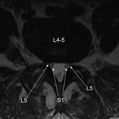
Bilateral L5 subarticular neural compression due to disc and facet disease.
Findings:
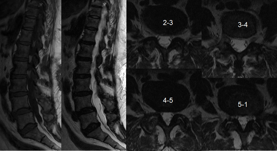
Sagittal MRI images demonstrate Multilevel disc height loss and desiccation with mild osteophyte formation. Grade 1 anterolisthesis is present at L4-5 measuring less than 4 mm. Axial images at selected levels demonstrate asymmetric subarticular narrowing at L4-5 causing compression of the bilateral L5 nerve roots which is delineated by the arrows on the final image, caused by facet hypertrophic changes asymmetric on the right. A diffuse disc bulge contributes to the subarticular narrowing at L4-5.
Discussion/Differential Diagnosis:
BACK TO
MAIN PAGE