Neuropathic Spondyloarthropathy
Findings:
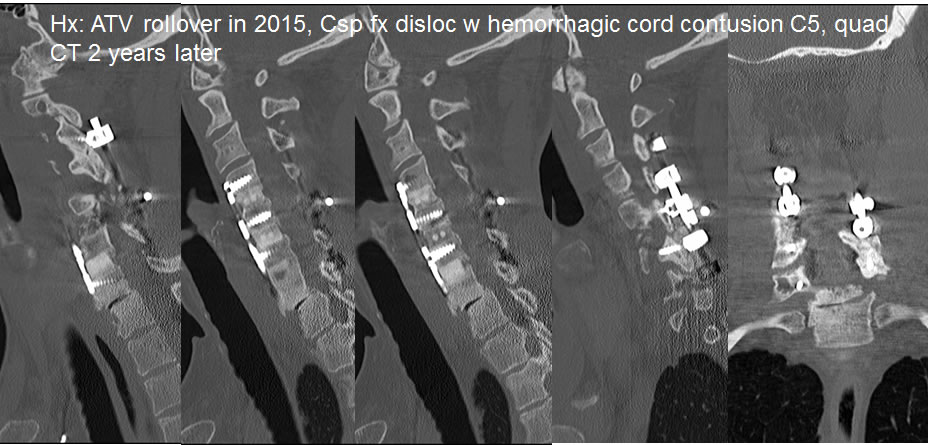
Sagittal images of the cervical spine performed two years after cervical spine trauma demonstrate well incorporated anterior fusion extending from C4 through C7. There is asymmetric marked disc space narrowing at C7-T1 with vacuum phenomenon, extensive sclerosis, and mild osteophyte formation, associated with mild to moderate anterior subluxation.
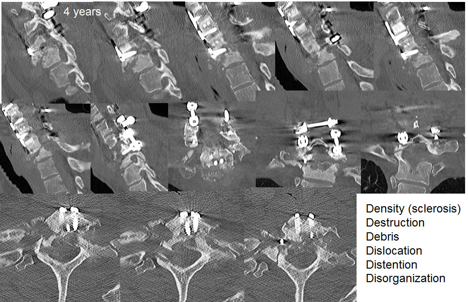
The follow-up CT images performed two years later demonstrate development of destructive changes within C7 and T1, loosening of the C7 screws, and progressive anterior subluxation. Right laminar fracture is seen at T1. Extensive debris is present in the prevertebral and epidural space at this level. The imaging features of neuropathic joint disease are summarized.
Discussion/Differential Diagnosis:
BACK TO
MAIN PAGE