
Ruptured Dermoid
Findings:
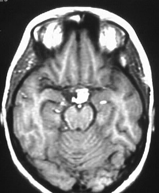
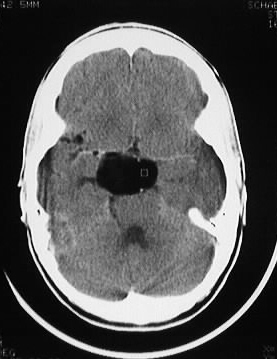
The axial CT image shows a fat density mass in the suprasellar
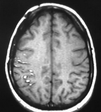
cistern. Axial noncontrast T1WI show nondependent layering hyperintense
material in the lateral ventricles and right cerebral sulci. This hyperintense
material exhibits chemical shift artifact characteristic of fat.
Discussion:
No real diferential diagnosis exists, since fat in the
vents is pathognomonic for a ruptured dermoid.
-ectodermal inclusion at neural tube closure
-epidermoid much more common
-40-50yr epidermoid, 20-30yr dermoid
-fat and calcification, no enhancement unless
infected, +/- fat/fluid level
-dermoid may have sinus tract if located
superficially-->recurrent meningitis, infection
-chemical meningitis when ruptured