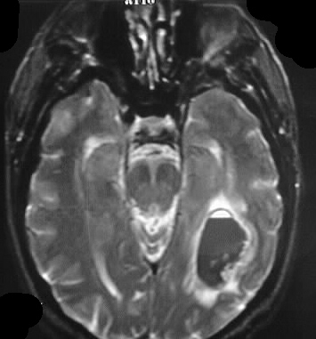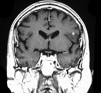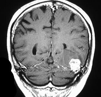


Hemorrhagic Metastatic Choriocarcinoma
Findings:
Large inhomogenous lesion left temporoparietal region
with surrounding edema, fluid-fluid levels on T2 (hemorrhage), nodular
enhancement. 2nd focus of nodular enhancement in left frontal operculum
region.
Differential Diagnosis:
hemorrhagic mets: lung, melanoma, thyroid, renal, chorio.
-illustrates importance of contrast enhancement when evaluating parenchymal hemorrhage. If only one lesion were present, primary brain tumor (GBM) would be most likely consideration.