Findings:
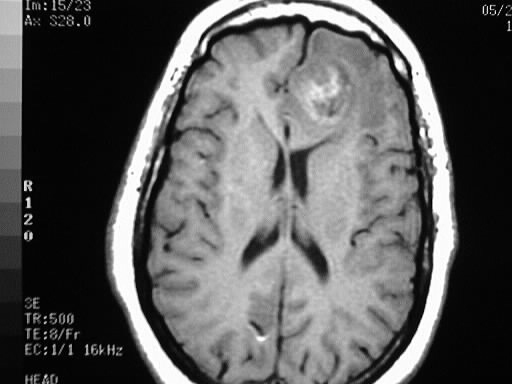
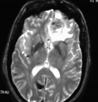
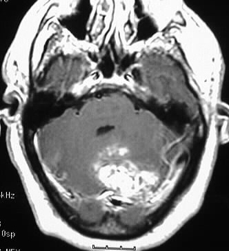
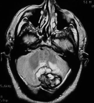
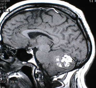
Large masses are present in the left frontal lobe
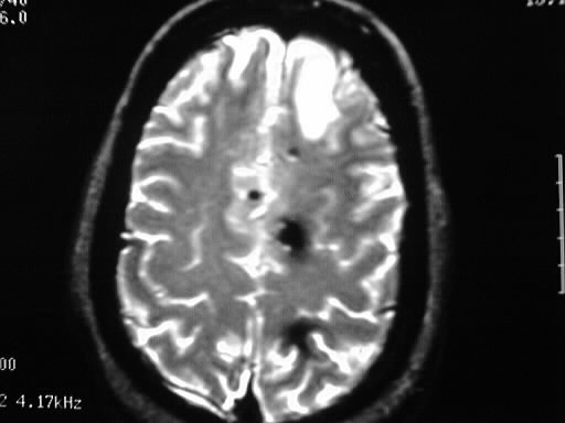
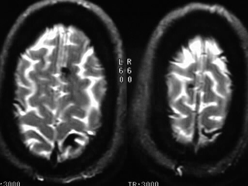
and midline posterior cerebellum, with smaller multifocal lesions in the
high gray/white junction. The larger lesions show nodular enhancement and
hyperintensity on T1WI. The smaller lesions show marked blooming on T2WI
indicative of blood products.
Discussion:
-hyperintensity
on T1 in this case= combination of hemorrhage (methemoglobin) and/or melanin
-ddx:
-hemorrhagic mets (melanoma, thyroid, renal, lung etc.)
-multiple cavernomas (usually no edema or masslike enhancement)