Dural AV fistula
Findings:
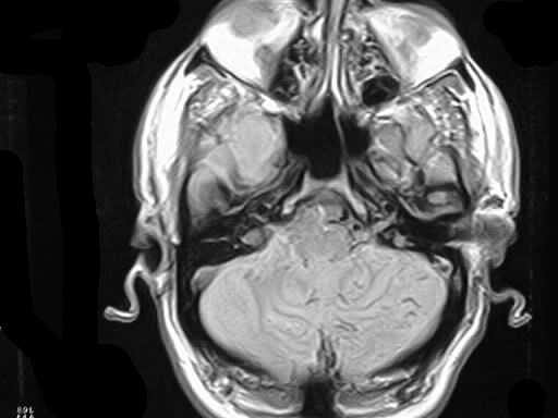
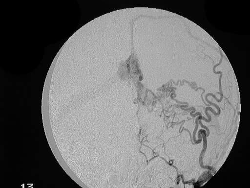
Axial PDWI shows abnormal flow voids along pial surface
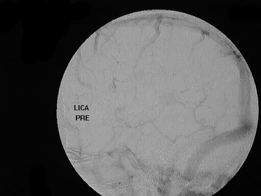
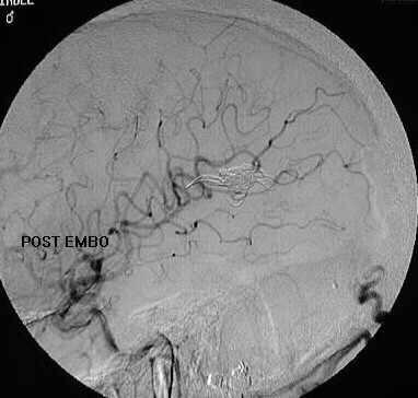
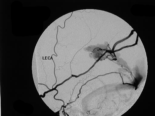
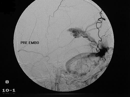
of cerebellum. DSA shows vascular malformation fed by ECA branches and
draining in region of Vein of Galen/ straight sinus via pial veins over
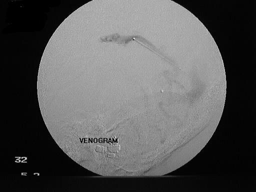
cerebellar hemisphere. Transvenous coil embolization performed.
Discussion:
This type of dural AV fistula is at high risk for hemorrhage
due to pial venous drainage. Inferior frontal dural AVFs are also at high
risk for hemorrhage.