Carotid Cavernous Fistula (ruptured cavernous carotid aneurysm)
Findings:
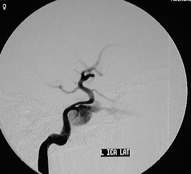
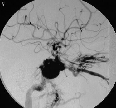
Lateral left ICA injection shows early filling of a large
saccular space, arising from the cavernous carotid. The second frame of
the injection shows rapid early filling of numerous veins, including a
markedly dilated superior ophthalmic vein, pterygoid plexus, and cavernous
sinus.
Differential Diagnosis:
trauma (basilar skull fx), ruptured cavernous sinus AVM,
ruptured cavernous carotid aneurysm.
Discussion:
Trauma is the most common cause of carotid-cavernous
fistula, due to skull base fracture with shear force against fracture fragments.
Clinically, patients present with chemosis, proptosis, bruit, and possible
visual loss. Spontaneous fistulas may be caused by atherosclerotic disease
and/or ruptured ICA aneurysm. Dural AVF may also be seen in this location,
but these are lower flow lesions and may be less symptomatic. These high
output fistulas may be occluded with balloons placed in the cavernous sinus.
If there is adequate collateral circulation across ACOM, the ICA may be
occluded by placing balloons distal and proximal to the fistula.