Arteriovenous Malformation
Findings:
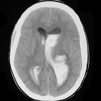
Axial CT image shows a large amount of hemorrhage
in mildly dilated lateral ventricles, associated with parenchymal hemorrhage/hyperdensity
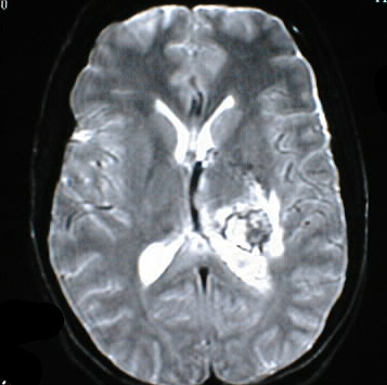
in the left thalamus. The axial MR images show various ages of blood products
in the posterior left thalamus, asociated with surrounding T2 hyperintensity
representing edema. A large flow void is present in the region of the left
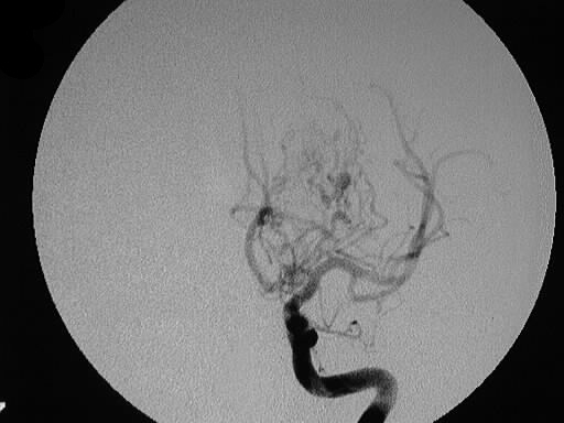
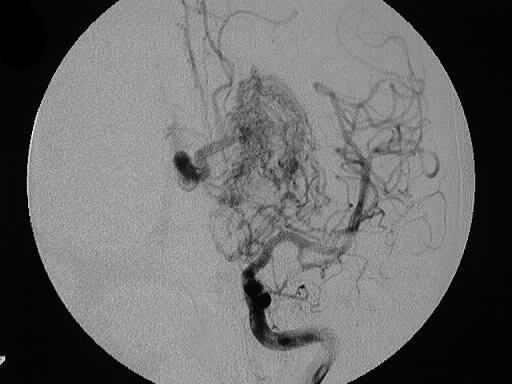
internal cerebral vein. AP early phase left internal carotid arteriogram
shows a complex tangle of vessels in the region of the thalamus, associated
with a large early draining vein.
Differential Diagnosis:
For intraventricular hemorrhage in this location seen
on CT, the differential diagnosis would include hypertensive hemorrhage
with rupture into the ventricles, hemorrhagic tumor (primary or met), or
ruptured vascular malformation. No other reasonable ddx for MR and angio
appearance except for the unlikely highly vascular glioma.
Discussion:
-5% of intracranial hemorrhages from vascular
malformation (includes cavernoma and others)
-80-90% supratentorial, 2-3%/yr risk of
bleed- 2x-3x increased in year following bleed
-types:
-pial (75%)-
ICA supply only
-pial/dural
(15%)- ICA/ECA supply
-dural (10%)-
ECA supply, acquired (dural sinus thrombosis)
-graded according to size, location, and
vascular supply (deeper, larger, multiple supply/drainage= higher grading)
-replace normal brain- usually solitary
(multiple in Wyburg -Mason and Osler Weber Rendu)
-clinical presentation: hemorrhage 50%,
sz 25%, neuro deficit 20-25%.
-+/- ring enhancing on CT, 25% Ca++, 10%
feeding artery aneurysm
This lesion represents a pial AVM, and would be difficult
to treat surgically due to its deep location and deep venous drainage.