R MCA Distribution Infarction
Findings:
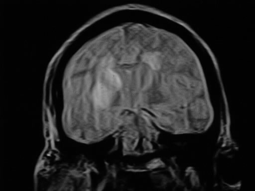
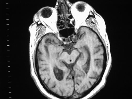
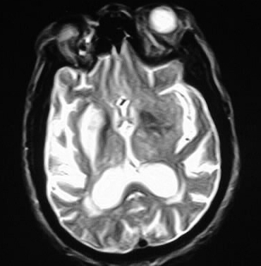
Axial and coronal MR images show abnormal signal in the
right temporal lobe and right basal ganglia,with sparing of the thalamus.
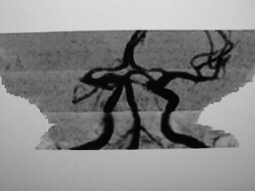
TOF MRA image shows abrupt termination of flow signal in the right M1 segment,
consistent with occlusion.
Discussion:
Ischemic Stroke
-no clear source in 25%
-if source identified- 2/3 thrombotic, 1/3 embolic
-20% lacunar- small vessel thrombotic
-other causes (5%): vasculitis, vasospasm, coagulopathy,
global hypoperfusion, venous thrombosis
-children: CHD, infection, trauma
-young adults: cardiac, drugs, dissection
-elderly: atherosclerosis, amyloid, coagulopathy
-brain gets 20% of CO, autoregulated
-GM 3-4x WM
-normal 55cc/100g/min- injury if <20cc
-<22- reversible
injury
-<18- loss of
action potential
-<12- 2 hrs-
cell death
-vulnerability: purkinje cells, hippocampus >>
glial cells and endothelium
-imaging
-cytotoxic
edema- begins 6h, peaks at 3-7 days. transudate, swelling without signal
change
-vasogenic
edema- begins 3-8 hrs, signal change
-diffusion
imaging- changes in 2 min- <15-20cc/min
-contrast
enhancement
-CT gyral at 7d-->peak 7-14d-->up to 3 months
-MR early intravascular enhancement, then parenchymal enhancement at 3d-3wks,
resolves 3 months