Cerebral Abscess
Findings:
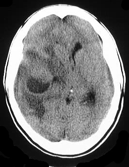
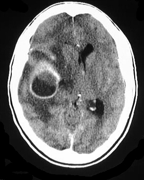
Axial noncontrast and contrast CT
images show a large ring enhancing mass involving the right frontal lobe,
with internal low density and nondependent debris, associated with moderate
surrounding edema and mass effect. There is moderate right to left midline
shift.
Differential diagnosis:
Glioma, metastasis, abscess, hematoma
(less likely). Helpful distinguishing features: thin uniform wall with
+/- mesial thinning.
Discussion:
-direct vs. hematogenous(most common),
anaerobes overall most common but may be S. aureus after surgery or trauma.
-frontal and parietal lobes
-lethargy, nausea, vomiting, fever(<
50%), meningeal signs(< 30%)
-rapid progression helps distinguish
from tumor
Stages:
1-early cerebritis 3-5 d
-poorly defined
area of necrosis- may be nl CT or hypodense, mild mass effect and patchy
enhancement. MR nonspec hypo/hyper.
2-late cerebritis 1-2 wks
-thick, irregular
wall enhancement with vasogenic edema. may have delayed fill in, may respond
to Abx.
3-early capsule
-well defined rim
enhancement, low T2.
4-late capsule
w
-better defined
rim with possible loculation, paramagnetic effect, possible mesial thinning.
BACK TO MAIN PAGE