Multiple Cavernomas
Findings:
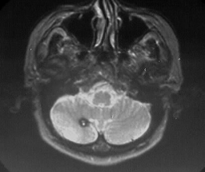
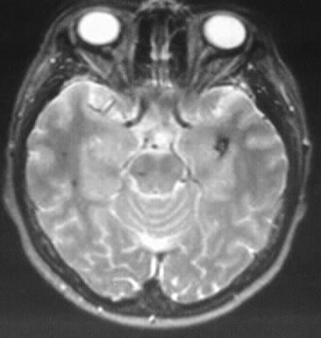
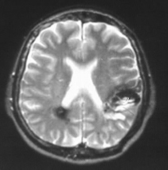
Axial T2WI show numerous hypointense lesions with hyperintense/heterogenous
centers, the largest of which is in the posterior left frontal lobe.
Differential Diagnosis:
cavernomas, multiple shear injury/contusions (lessl likely
due to location), hemorrhagic metastases
Discussion:
-hemorrhage risk 0.5-1%/yr/lesion
-"popcorn" appearance is characteristic, commonly coexistent
DVA
-thin walled sinusoidal vessels, usual normal CT/angio
-reticulated enhancing heterogenous lesion on MR with
hemosiderin rim (blooming artifact)