Aggressive Pineocytoma
Findings:
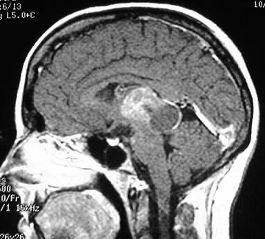
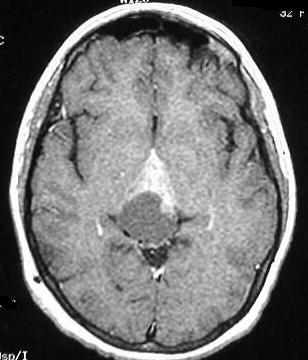
Sagittal and axial T1WI postcontrast show a mass in the
region of the pineal gland with an enhancing portion extending into the
posterior third ventricle and possibly invading the midbrain/thalamus.
The mass has a rounded nonenhancing portion posteriorly within the QP cistern.
Differential Diagnosis:
Germ cell tumor, pineal cell tumor (pineoblastoma, pineocytoma),
glioma, met. Location above the tectum and below the Vein of Galen localizes
this mass to the pineal region.
Discussion:
Pineal region masses may present with Parinaud syndrome
(upward gaze palsy), precocious puberty, or increased intracranial pressure.
60% are germ cell neoplasms (germinoma, seminoma, embryonal etc.). A calcified
pineal mass is more likely to be germinoma in males and pineocytoma in
females. Imaging usually can't distinguish between germ cell and pineal
cell tumors.
-germinoma (most common germ cell tumor)- 60%
-peak incidence puberty, common CSF
dissemination
-iso/hyper CT, hypo/hyper MR
->50% fysr (radiosensitive)
-pineocytoma/pineoblastoma- 15%
-children: pineoblastoma -similar to medulloblastoma
-rare "trilateral retinoblastoma"
-adults- pineocytoma- less CSF spread
-iso/hyper CT, iso/hyper MR, intense enhancement
-others: gliomas, mening, arach cyst, vein of Galen
aneurysm, lipoma, pineal cyst- 25%