
Trigeminal Schwannoma
Findings:
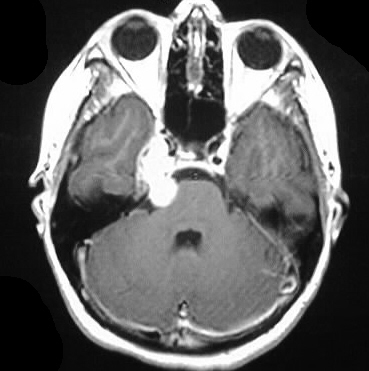
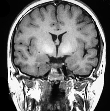
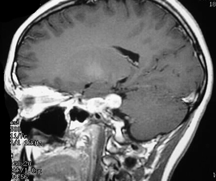
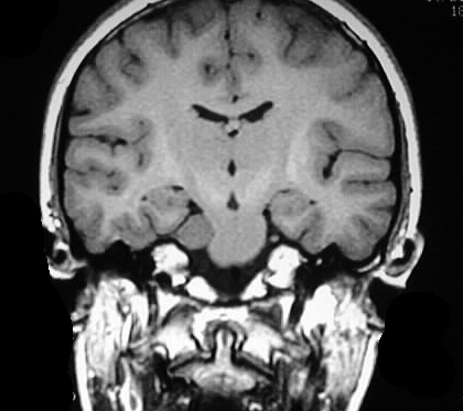
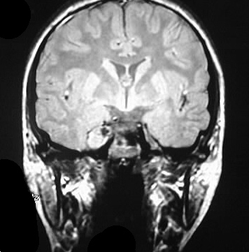
Multiplanar T1 pre/postcontrast images and coronal PDWI
show a strongly enhancing mass that occupies the right Meckel's cave region
and causes mild mass efect on the adjacent temporal lobe and pons. The
lesion is bilobed and extends into the superior portion of the CP angle/pontine
cistern. The mass is isointense to gray matter on T1 and hyperintense on
PD.
Differential Diagnosis:
trigeminal schwannoma, meningioma, metastasis, less likely
chondrosarcoma. Hyperintensity on PD makes meningioma less likely.
Discussion:
-schwannoma=8-10% of all intracranial
neoplasms, 60-90% of CPA mases
-VIII and V origin most common, +/-encapsulated
-present age 40-60 with neuralgia,
masticator weakness. F>M 2:1.
-mostly sporadic but 5% inherited
(MISME)
-imaging: hypo/iso on CT +/- Ca++,
hypo or iso T1, hyper T2
-most commonly solid but may be cystic,
necrotic, hemorrhagic