Craniopharyngioma
Findings:
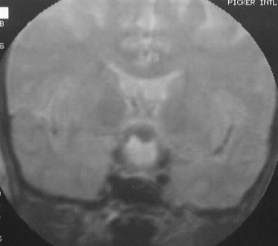
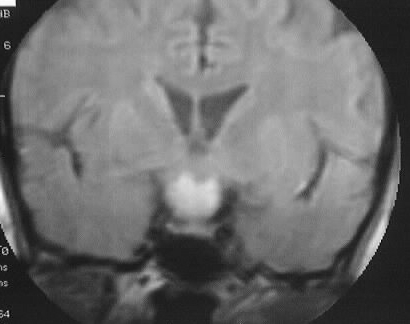
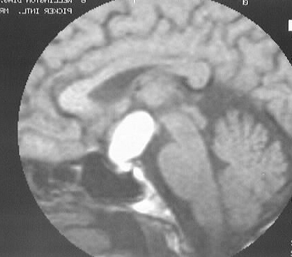
Sagittal T1, coronal T2, and coronal T1WI show a suprasellar
mass with hyperintensity on both T1 and T2WI. The mass is predominantly
suprasellar, with extension into the sella.
Differential Diagnosis:
craniopharyngioma, rathke's cleft cyst, hemorrhagic adenoma
less likely.
Discussion:
-present clinically with increased intracranial pressure,
hypothalamic dysfunction, or chiasmatic compression symptoms
-origin: cranio from pars tuberalis, Rathke's from pars
intermedia
-location: 70% intra and suprasellar, 10% suprasellar
only, 10% intrasellar only
-age peaks: 5-10 and less so at 50-60
-most common suprasellar mass in children (50%)
-imaging: 80% Ca++ in children, 40% Ca++ adults, 90%
cystic, 90% rim enhance, hyper T1/T2 (cholesterol), +/- optic tract signal
abn
-main ddx is Rathke's cleft cyst (RCC enhances minimally
or not at all)