Dural Fibrosis after Shunt Placement
Findings:
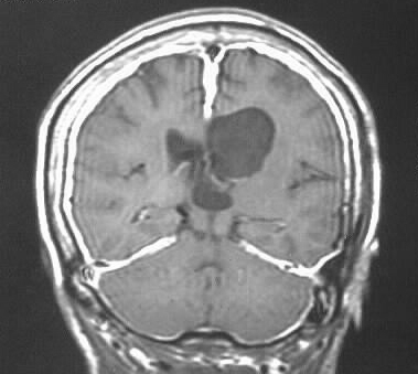
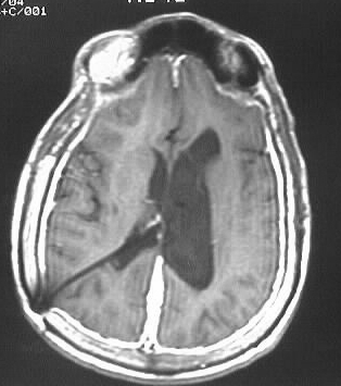
Coronal and axial postcontrast T1WI show uniformly thickened
enhancing dura, without nodularity. The leptomeninges are spared. Shunt
tubing enters from the right. The lateral ventricles are asymmetric.
Differential Diagnosis/Discussion:
Dural fibrosis and intracranial hypotension would be
difficult to distinguish, but the prominence of the left lateral ventricle
argues against overshunting. Metastatic disease and inflammatory causes
such as sarcoidosis could also be considered, but this uniform pattern
would be atypical.