Acute Disseminated Encephalomyelitis
Findings:
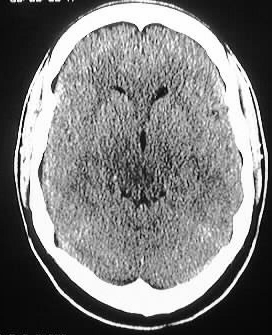
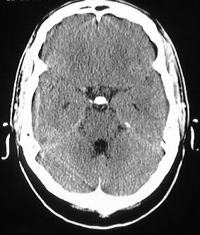
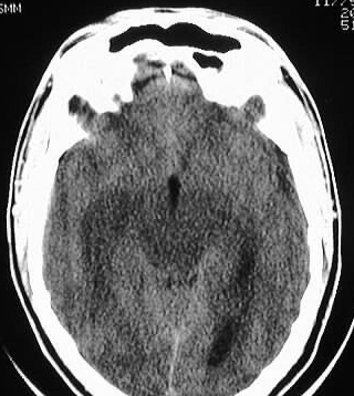
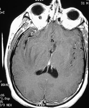
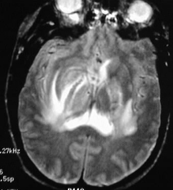
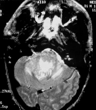
Axial CT images show patchy areas of low atttenuation
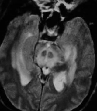
involving the midbrain and periventricular white matter. Axial T1 postcontrast
and T2WI show extensive signal abnormalities in the basal ganglia, midbrain,
and posterior fossa structures. Minimal spotty enhancement is present.
Differential Diagnosis:
ADEM, MS, PML, SSPE, also mesorhombencephalitis as seen
with listeria, herpes, Behcet's, sarcoid. Multifocal glioma could have
a similar appearance.
Discussion:
ADEM usually occurs in children within weeks following
viral illness or vaccination, and likely represents an autoimmune response
to myelin. Onset is abrupt and monophasic to distinguish from MS. Many
completely resolve over weeks, but there is a 20% rate of permanent deficit
and 15-20% mortality. The disease responds to steroids. Imaging features
may resemble MS but are usually more extensive, with involvement of subcortical
white matter, brainstem, cerebellum, spinal cord, and possibly basal ganglia.
Enhancement is variable (nodular, gyriform, patchy, ring, etc.). There
is typically no mass effect.