Epidural Hematoma
Findings:
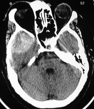
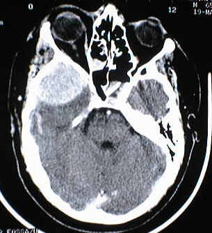
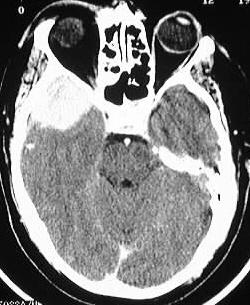
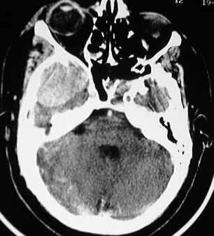
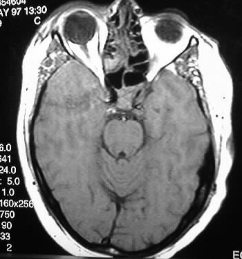
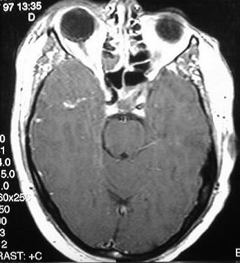
Axial noncontrast and contrast enhanced CT images show
a well defined hyperdense extraaxial collection in the right middle cranial
fossa. Te enhanced CT images show increased hyperdensity of the collection
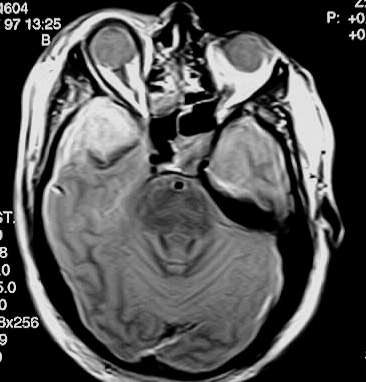
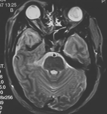
consistent with clotting. The collection is isointense on T1, hyperintense
on T2WI and does not enhance. The signal characteristics are consistent
with acute hemorrhage, with some dependent deoxyhemoglobin seen on the
T2WI.
Discussion:
MR and CECT are rarely performed in the setting of acute
trauma, but were performed in this case due to an inconsistent clinical
picture. These images nicely demonstrate the MR signal characteristics
of blood products in the acute stage.
MR of hemorrhage:
-oxy- diamagnetic-
T2 hyper 1st echo, T1 hypo/iso- difficult to dist from CSF in SAH
(iso/bright)
-deoxy- paramagnetic-
accelerated dephasing- T2 hypo, T1 hypo/iso (iso/dark)
-met- intracell-
local field gradients btw cell and protons outside- sig loss on T2 (bright/dark)
-met- extracellular-
gradients lost- T2 now similar to CSF (bright/bright)
-hemosiderin-
paramagnetic- hypo T2 (dark/dark)