
RICA Dissection with acute R MCA distribution infarction
Findings:
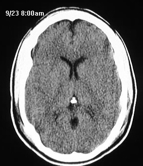
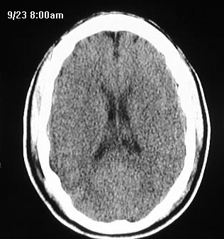
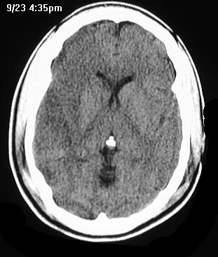
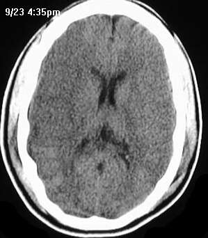
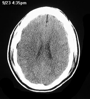
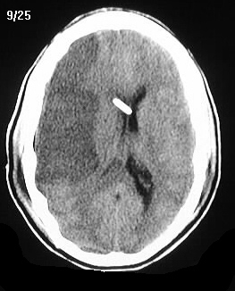
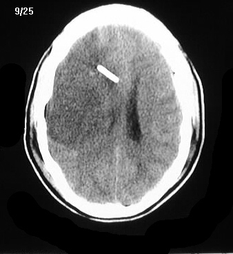
Serial CT images show developing hypodensity and mass
effect in the right cerebral hemisphere, with sparing of the medial frontal
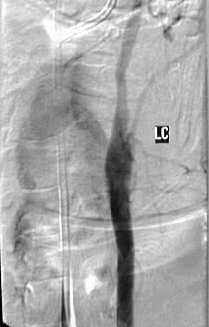
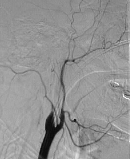
and occipital lobes. A ventricular shunt was placed. LCCA arteriogram shows
minimal luminal irregularity and tapering of the left ICA. RCCA arteriogram
shows a tapered abrupt occlusion of the right ICA.
Discussion:
Evolving infarct in a trauma patient should be investigated
for the possibility of dissection. Occasionally, vascular dissection may
occur with relatively minor trauma to the neck, abrupt head movement, or
chiropractic manipulation. Underlying vessel abnormalities such as kinking,
FMD, Marfan's disease, and long standing hypertension may predispose to
dissection. Dissections may be identified on MR as a characteristic high
signal rim surrounding the ICA flow void at the skull base. Conventional
angiography is best to define the initial extent of vessel abnormalities,
but MRA may be adequate for follow up studies. Strokes are usually caused
by distal emboli rather than occlusion, unlike this case. Most dissections
either remain the same or resolve on follow up examination.