Diffuse Axonal Injury
Findings:
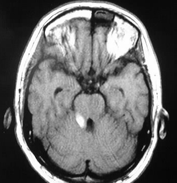
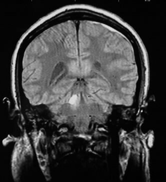
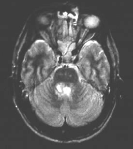
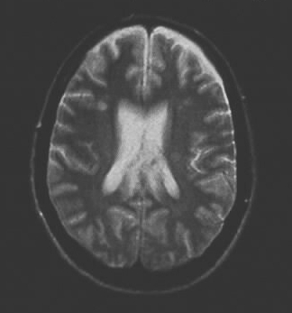
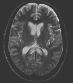
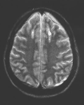
T1 and T2 weighted images show extensive areas of abnormal signal involving
the right superior cerebellar peduncle, bilateral splenium of corpus callosum,
and bilateral high frontal gray/white junction. Small bifrontal extraaxial
fluid collections are present as well, consistent with subdural hygromas
or aging subdural hematomas. A small focus of hemorrhage is evident in
the left sylvian fissure as well.
Differential Diagnosis:
The constellation of findings is highly consistent with
traumatic injuries, with DAI. Isolated abnormal signal in the corpus callosum
could rarely be due to tumor, but it would not be this symmetric.
Discussion:
Most patients with significant shear injury are comatose
from the point of impact, and the degree of injury may be inapparent on
initial head CT. The mechanism of DAI involves rapid accleration/deceleration
forces with disruption of axons. DAI is responsible for the majority of
coma and other poor outcome associated with closed head trauma. Common
locations of shear injury include corpus callosum splenium, gray/white
junction, and dorsolateral brainstem. MR is more sensitive for demonstration
of tiny signal abnormalities. Imaging findings include focal hyperintensities
+/- blood products, for which gradient echo sequences may be more sensitive.
CT findings may be normal if nonhemorrhagic shear is involved, but hemorrhagic
shear injury may have central hyperdensity with peripheral edema.