
Hemangioblastoma
Findings:
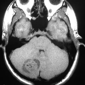
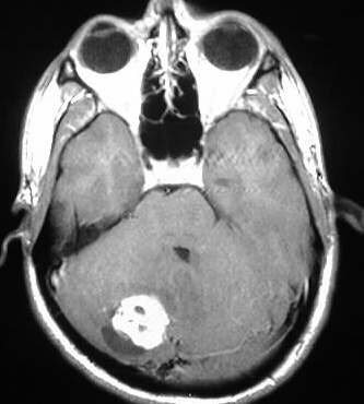
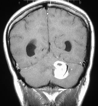
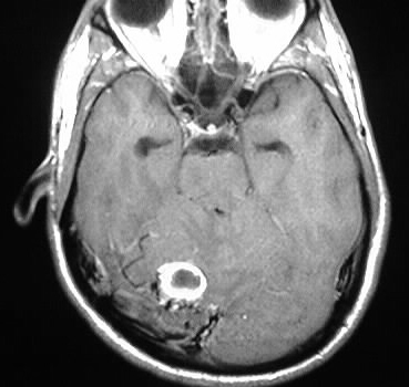
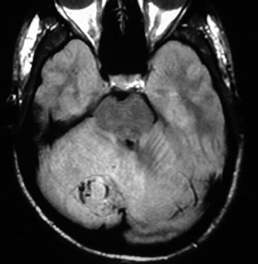
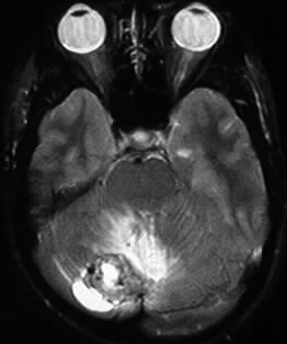
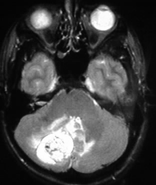
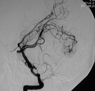
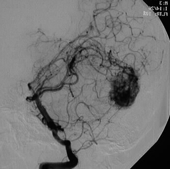
Multiple MR images and lateral vertebral DSA show a strongly
enhancing hypervascular mass in the right cerebellum, asociated with a
small cystic component and surrounding signal abnormality extending throughout
the right cerebellar hemisphere. The mass is isointense on T1 and mostly
hyperintense on T2.
Differential Diagnosis:
hemangioblastoma, metastasis, glioma. A flow void within
the nodule is a key distinguishing point.
Discussion:
Hemangioblastoma is a benign tumor of endothelial origin,
seen in young-middle age, and is the most common primary intraaxial
tumor of posterior fossa in adults. It represents 1-2% of intracranial
neoplasms and 10% of posterior fossa tumors.
-4-20% assd. with VHL. 40% of multiple tumors
assd. with VHL.
-45% of VHL pts. develop hemangioblastoma
-cerebellar hemispheres(80%), cord (10%),
medulla (2%)
-no capsule, pial supply (superficial),
rare calcification
-60% cyst/nodule, 40% solid
-iso T1, hyper T2, edema. flow voids in
nodule, ill defined +/- hemorrhage
-cyst portion may have higher T1/T2 signal
than CSF due to protein
-10-40% assd with polycythemia (epo)
reference: Osborn, A.; Tong, K. Handbook of Neuroradiology: Brain and Skull. 2nd ed. 1996: Mosby Year Book. pp. 296-297.