Cerebral Amyloid Angiopathy (autopsy proven)
Findings:
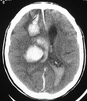
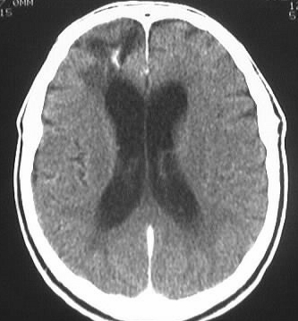
Axial CT images performed on different days show a streaklike
hyperdensity in the right frontal lobe with surrounding low attenuation,
which develops into a large parenchymal hemorrhage with rupture into the
lateral ventricles. A second focus is apparent in the right corona radiata
region.
Differential Diagnosis:
hemorrhagic infarction, amyloid angiopathy, less likely
hypertensive hemorrhage, trauma
Discussion:
Amyloid angiopathy is a less considered but common cause
of parenchymal hemorrhages in the normotensive elderly. Pathologically,
amyloid deposits in the media and adventitia of medium size and small cortical
leptomeningeal vessels, causing vascular thickening and fragility. The
condition is not associated with systemic amyloidosis. Approximately 30%
of cases are associated with progressive dementia. The prevalence increases
with advancing age, with a large number occurring over age 90. Imaging
features include superficial infarcts and hemorrhages of varying ages,
with a predilection for the corticomedullary junction and parietooccipital
regions. The basal ganglia are usually spared.