Glioblastoma Multiforme
Findings:
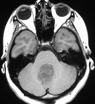
Axial T1WI shows a hypointense mass in the region of
the fourth ventricle with ill defined posterior demarcation. The mass displaces
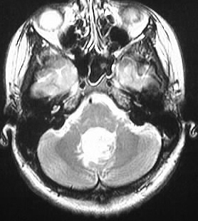
the brainstem anteriorly. The mass is markedly hyperintense on the axial
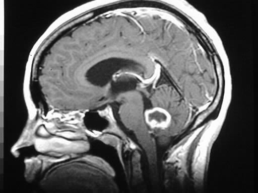
T2WI and there is surrounding signal abnormality in the cerebellum. A thick,
irregular rim of enhancement is present on the sagittal T1 postcontrast
image.
Differential Diagnosis:
The enhancement characteristics are not consistent with
a medulloblastoma, pilocytic astrocytoma, or hemangioblastoma. Ependymoma
and metastasis could be considered, with abscess much less likely. Unfortunately,
I don't know the age of this patient, which would help guide the differential
diagnosis.
Discussion:
This mass appears to be arising from the posterior fourth
ventricle. Intraventricular GBMs are uncommon but not rare. GBM is the
most common glioma, with an age peak of 45-55, M>F. Microscopically wide
invasion is the rule despite possibly having a well circumscribed appearance.
GBM may have occasional calcification and may be markedly hypervascular
with flow voids, simulating AVM on angio. The tumor may become more necrotic
and calcified with treatment. Prognosis is dismal, with a median survival
measured in months.