Craniopharyngioma
Findings:
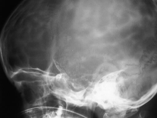
The lateral skull film shows marked expansion of the
sella and a large faint rimlike area of calcification projecting over the
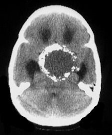
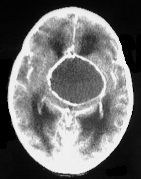
suprasellar region. Axial CT images show a large sellar region mass with
internal low density and thick rimlike calcification, associated with hydrocephalus. No central enhancement is present.
Differential Diagnosis:
This constellation of findings is highly consistent with
craniopharyngioma, and this is by far the most likely diagnosis. Germ cell
tumor could be considered if there were more enhancing solid component,
and teratoma could be considered also but would be less likely.
Discussion:
-present clinically with increased intracranial pressure,
hypothalamic dysfunction, or chiasmatic compression symptoms
-origin: cranio from pars tuberalis, Rathke's from pars
intermedia
-location: 70% intra and suprasellar, 10% suprasellar
only, 10% intrasellar only
-age peaks: 5-10 and less so at 50-60
-most common suprasellar mass in children (50%)
-imaging: 80% Ca++ in children, 40% Ca++ adults, 90%
cystic, 90% rim enhance, hyper T1/T2 (cholesterol), +/- optic tract signal
abn
-main ddx is Rathke's cleft cyst (RCC enhances minimally
or not at all)