Laryngocele
Findings:
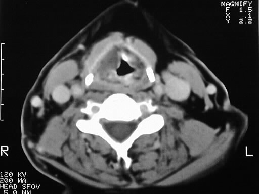
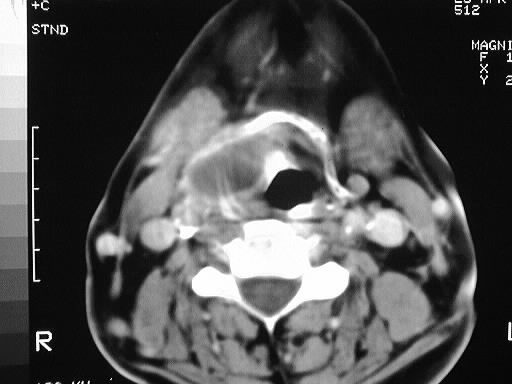
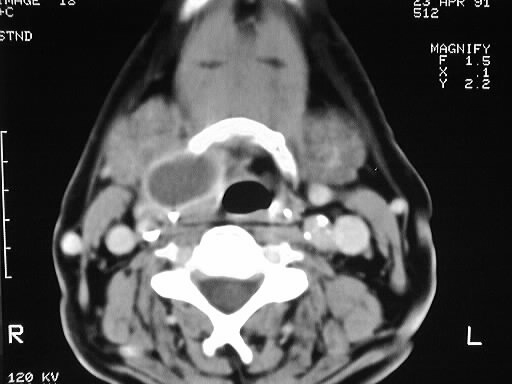
Axial contrast CT of the neck shows a fluid density mass
arising at the level of the left false cords, extending superiorly to the
level of the hyoid bone. The lesion has a thin enhancing rim.
Differential Diagnosis:
laryngocele, laryngopyocele, abscess, less likely dermoid.
thyroglossal duct cyst is not likely due to location deep to the strap
muscles.
Discussion:
Laryngocele develops as a consequence of chronically
increased intraglottic pressure as may be seen in musicians, glass blowers,
or excessive coughing. The lesions represent a dilated appendix of the
laryngeal ventricle. Laryngoceles are classified as internal, external,
or mixed, according to their relation to the thyrohyoid membrane. Mixed
types are the most common. The internal type, as in this case, may cause
stridor. The lesions may become infected as well (see unknown # 81). Laryngoceles
that develop without a known predisposing factor should raise the suspicion
of an underlying neoplasm obstructing the laryngeal ventricle.