Findings:
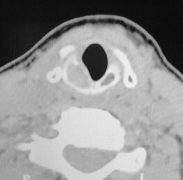
Axial noncontrast CT at the level of the cricoid cartilage
shows expansion, soft tissue replacement, and anterior disruption of the
right cricoid, associated with mild effacement of the posterolateral airway.
A tiny hyperdensity is present in the center of this mass, most likely
representing matrix calcification.
Differential Diagnosis:
Anterior disruption of the cartilage border favors malignancy,
of which chondrosarcoma would be the most common in this location and appearance.
Other considerations include metastasis, local invasion from adjacent squamous
cell carcinoma, or less likely lymphoma.
Discussion:
Calcifications in typical ringlike or arclike patterns
may give a clue to the specific histologic diagnosis, but in the absence
of this the distinction between benign and malignant processes may be difficult.