Findings:
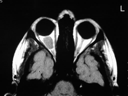
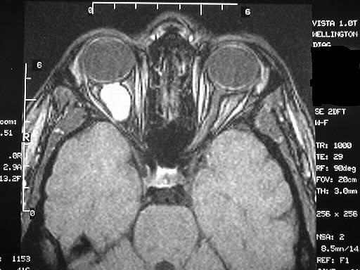
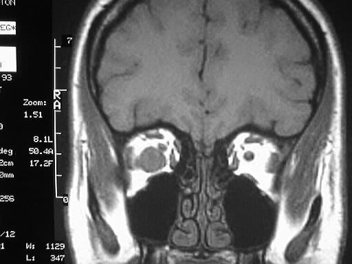
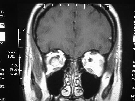
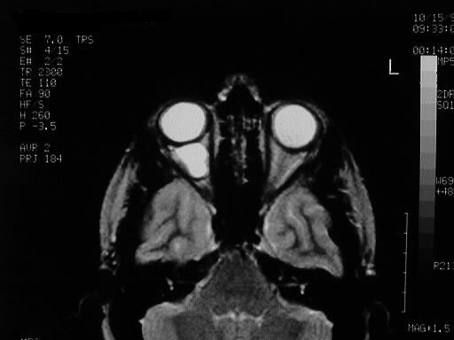
Multiple MR images show a right intraconal mass which
is isointense to grey matter on T1, markedly hyperintense on T2, and enhances brightly.
The right optic nerve is seen as a separate structure, displaced superomedially
by the mass.
Differential diagnosis:
hemangioma, lymphoma, metastasis, fibrous histiocytoma,
schwannoma. Hemangioma is most likely given the signal characteristics.
Discussion:
Cavernous hemangioma is the most common primary intraconal
mass in adults.
-benign noninfiltrative hamartoma, slowly
enlarging
-25-70, peak 25-40
-hyperdense on CT, intense enhancement,
+/- phleboliths, occ hemorrhage
-iso T1/hyper T2