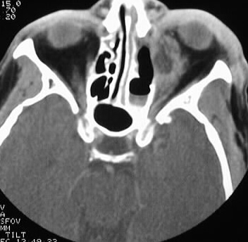
Findings:
Axial CT of the orbits with contrast shows a small superiosteal
fluid collection in the left orbit, associated with focal defects in the
medial orbital wall and adjacent ethmoid sinus opacification. Moderate
thickening of the left medial rectus muscle is present as well.
Differential Diagnosis:
This almost certainly represents an infectious process,
but the appearance is not organism specific. The presence of bone destruction
indicates an aggressive infection, such as mucormycosis. Bacterial orbital
infection is relatively more common.
Discussion:
Infection is the most common orbital disease, representing
approximately 60% of cases. The most common bacterial organisms include
staph, strep, pneumococcus, and pseudomonas. Fungal disease such as aspergillus
and mucor may be seen in immunocompromised patients. Orbital cellulitis
is commonly caused by adjacent sinus disease. Without adequate treatment,
the inflammatory disease progresses from edema to subperiosteal phlegmon
and abscess to orbital cellulitis and abscess. Complications in the advanced
stages include cavernous sinus and ophthalmic vein thrombosis, visual loss,
opthalmoplegia, and intracranial abscess.
Mucormycosis is classified into rhinocerebral, pulmonary, gastrointestinal, and disseminated forms. The infection may be rapidly fatal if not treated surgically. Mucor is highly angioinvasive, which may cause brain infarction.