Amelanotic Melanoma
Findings:
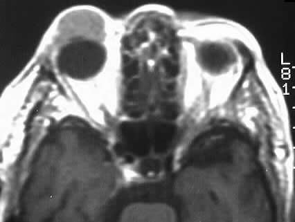
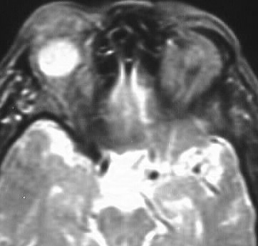
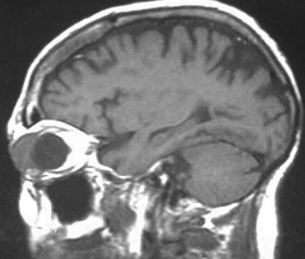
Multiple MR images show an extraconal mass in the right
orbit which has intermediate signal intensity on T2 and is isointense to
gray matter on unenhanced T1. The axial contrast enhanced T1 weighted image
shows mild homogenous enhancement of this lesion.
Differential Diagnosis:
This tumor has a nonspecific imaging appearance. Intermediate
signal on T2 suggests that this is a cellular lesion, and tumors such as
lymphoma could be considered. Other lesions to consider include metastasis
and amelanotic melanoma.
Discussion:
Melanin producing melanomas show hyperintensity on T1
and hypointensity on T2. Amelanotic lesions have a nonspecific appearance,
generally hypo/hyper T1/T2. Melanoma is the most common intraocular
malignancy in adults, but extraconal lesions are uncommon.