3rd Branchial Cleft Cyst (suspected)
Findings:
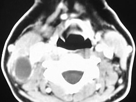
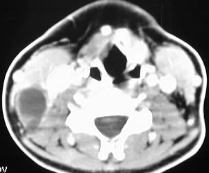
Axial neck CT with contrast demonstrates a fluid density
mass posteromedial to the right sternocleidomastoid muscle, associated
with a thin peripheral rim of enhancement.
Differential Diagnosis:
branchial cleft anomaly, necrotic lymph node, abscess,
lymphangioma
Discussion:
Of branchial cleft anomalies, those arising from the
2nd cleft are by far the most common. A 2nd branchial cleft cyst is situated
in the posterior submandibular space between the submandibular gland and
SCM muscle. 1st branchial cleft cysts are found in the region of the parotid
gland. 3rd branchial cleft cysts are found in the posterior cervical space,
as shown here. The cysts may be associated with a sinus tract or fistula,
and may present as recurrent infection.