

Delayed Presentation of LICA aneurysm
Findings:
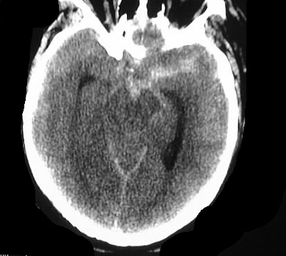
Axial noncontrast CT shows subarachnoid hemorrhage in
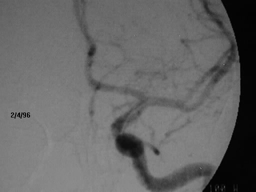
the basal cisterns with a left sided predominance. Initial LICA injection
shows no abnormality. A complete four vessel angiogram was performed which
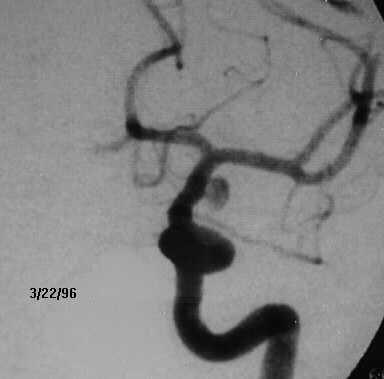
showed no abnormality (not shown). Follow up angiogram 6 weeks later shows
a cavernous internal carotid aneurysm.
Discussion:
Approximately 10-20% of angiograms in acute subarachnoid
hemorrhage are initially negative. Reasons for negative angiograms in this
setting include incomplete studies, errors in interpretation, and thrombosis
of the aneurysm. Only 10-25% of follow up angiograms show the aneurysm.
Other than trauma, benign perimesencephalic hemorrhage is the most common
cause of nonaneurysmal subarachnoid hemorrhage.