
Basilar Artery Aneurysm
Findings:
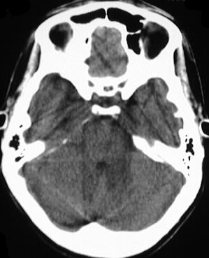
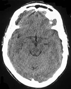
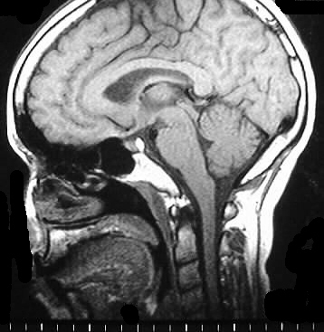
Axial noncontrast CT shows a small mass in the interpeduncular
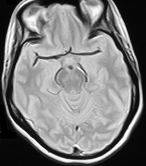
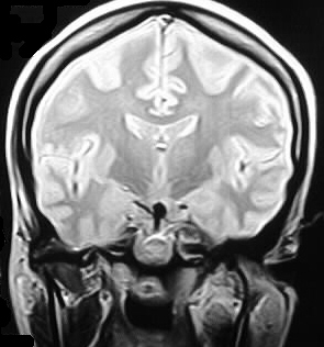
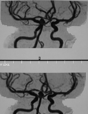
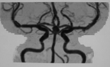
cistern region in the expected location of the distal basilar artery. MR,
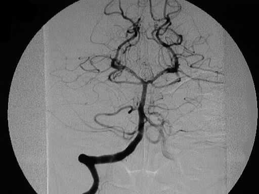
MRA, and arteriography show the aneurysm at the tip of the basilar artery.
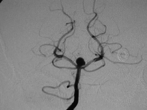
The final arteriographic image shows successful coil embolization of the
aneurysm.
Discussion:
Basilar artery aneurysms comprise approximately 10% af
all intracranial aneurysms. The pathologic lesion is reduction or absence
of the internal elastic lamina and/or media, either congenital or acquired,
which markedly reduces elastic tension and allows expansion of the vessel
diameter. 80-90% of nontraumatic subarachnoid hemorrhage is due to aneurysm.
The risk of bleeding is approximately 2.5% per year for lesions >6mm in
diameter, but any size aneurysm can bleed. Complications of SAH include
hydrocephalus, rebleeding, vasospasm (>7d) +/- infarct. 1/3 of patients
with ruptured aneurysm die immediately, 1/3 have long term disability,
and 1/3 are normal.
-grading SAH clinical: Hunt and Hess
-0- unruptured
-1- asympt or min H/A
-2- mod/severe H/A, nuchal rigidity, CN
palsy
-3- drowsiness, confusion, mild focal deficit
-4- stupor, mod/severe hemiparesis, early
decerebrate, vegetative
-5- deep coma, moribund
-+1 for vasospasm or systemic disease
-CT grading of SAH- Fisher
-0- unruptured
-1- no blood detected
-2- diffuse <1mm
-3- localized clot and/or vertical layer
>1mm
-4- intracerebral or IV clot with diffuse
or no SAH