Traumatic Anterior Cerebral Artery Pseudoaneurysm
Findings:
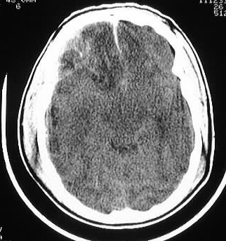
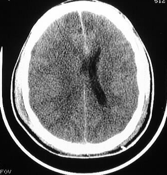
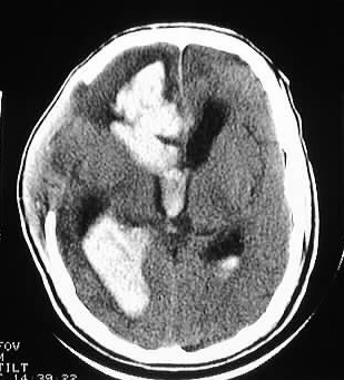
Axial CT images show numerous right frontotemporal contusions
with mass effect and effacement of the right lateral ventricle. A small
amount of pneumocephalus is present posteriorly. In addition, small amount
of subarachnoid hemorrhage is seen in the anterior interhemispheric fissure.
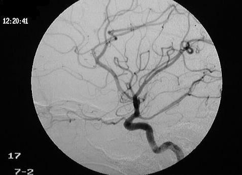
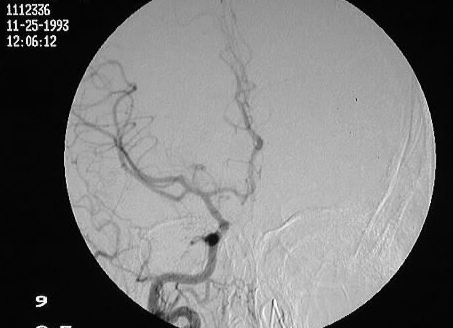
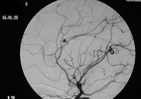
Arteriography was performed initially for suspected ICA injury, which was
negative. The patient developed severe headache 6 weeks later, at which
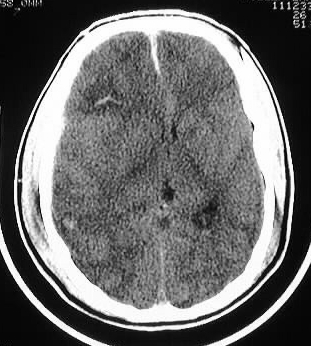
time an additional CT scan was performed. This study shows a large amount
of intraventricular hemorrhage, with a parenchymal hemorrhage in the right
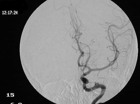
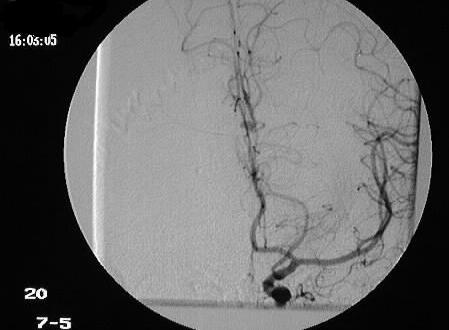
frontal lobe. Repeat cerebral arteriogram shows pooling of contrast in
a moderate size pericallosal artery pseudoaneurysm.
Discussion:
The anterior cerebral artery is susceptible to traumatic
injury due to increased shear forces along the genu of the corpus callosum.
Posttraumatic pseudoaneurysms of the posterior cerebral artery may occur
from impact on the tentorium. The intimal injury may not initially be apparent.
These lesions expand over time and are at a high risk for causing catastrophic
hemorrhage. In retrospect, the small amount of interhemispheric SAH may
have been a clue to the presence of arterial injury, but this could not
be assumed prospectively since small amounts of SAH are commonly seen in
trauma.