
Tuberous Sclerosis
Findings:
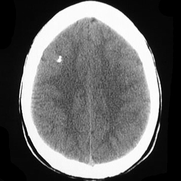
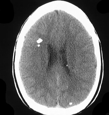
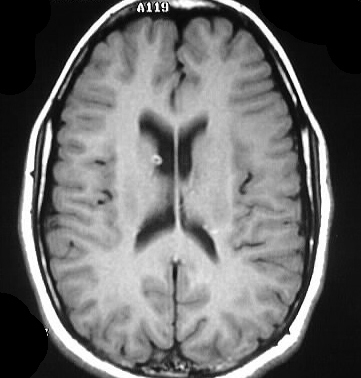
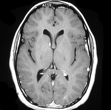
Axial CT images show chunky calcifications at the gray-white
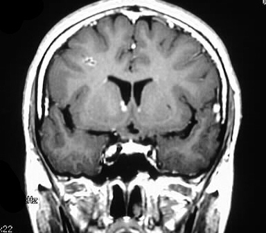
junction, without edema or mass effect. Multiple MR images show enhancement
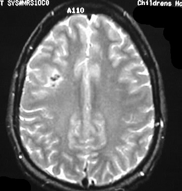
in the region of the right Foramen of Monro, with scattered areas of subcortical
T2 signal abnormality.
Discussion:
The constellation of findings is highly consistent with
a diagnosis of tuberous sclerosis. Clinically these patients present with
the triad of seizures (90%), mental retardation (50%), and adenoma sebaceum
(90%).
-autosomal dominant, 1/10,000-1/50,000
-chromosomes: TSC 1 9q, TSC 2 16p- forme fruste 5x more
common
-criteria (need 1)
-facial angiofibromas
-ungual fibroma
(17%)
-retinal hamartoma
-cortical
tubers (50%)
-subependymal
nodules
-multiple
renal AML
-presumptive (need 2)
-hypomelanotic
nodules
-shagreen
patch
-single AML
-multicystic
kidney
-cardiac rhabdomyoma
(30-50%)
-LAM pattern,
honeycomb lung
-first degree
relative with TS
-subependymal giant cell astrocytoma in 15% (WHO grade
I)
-other findings: retinal benign astrocytic hamartoma,
heterotopias, myelination disorder, ventriculomegaly