Cerebellopontine Angle Lipoma
Findings:
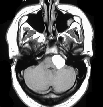
T1 unenhanced axial image shows a homogenous hyperintense
mass in the left CPA cistern, which exerts little mass effect on the pons.
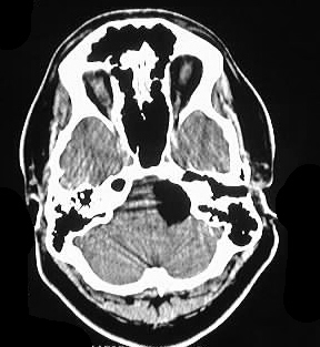
The fourth ventricle is normal in position. Axial CT images shows that
this mass is fat density. The mass remodels bone, indicating an indolent
process.
Differential Diagnosis:
Hyperintensity on T1 can be due to fat, methemoglobin,
melanin, enhancement, calcium, and proteinaceous fluid. Since the lesion
is clearly fat dansity on CT, lipoma is the only reasonable diagnosis.
Discussion:
CNS lipomas are congenital malformations which arise
from the meninx primitiva in formation of the subarachnoid cisterns, and
are associated with callosal and/or cerebral dysgenesis in over 50 percent
of cases. The lesions are also associated with facial anomalies and encephaloceles.
Lipomas are most commonly found in the dorsal pericallosal region. CPA
cistern lipomas are relatively uncommon.