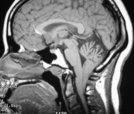
Chiari I Malformation
Findings:
Sagittal T1WI shows cerebellar tonsils well below the
foramen magnum, at least 2-3cm. The CSF space at the foramen magnum is
narrow. No syrinx is evident in the upper cervical cord. The fourth ventricle
is normal in size and configuration.
Differential Diagnosis:
None really appropriate for this case except Chiari I
malformation. Tonsillar herniation could be seen in a patient with markedly
increased intracranial pressure, but there is no evidence of that in this
case.
Discussion:
3mm below ="mild tonsillar ectopia"- no clinical significance,
3-5mm gray area, >5mm= Chiari I- may have cranial neuropathy due to brainstem
compression, central cord syndrome due to syrinx.
Chiari I is by far the most common of the Chiari malformations. Etiologic theories include embyologic anomaly of craniocervical junction, intrauterine tonsillar herniation due to hydrocephalus, or acquired deformity from platybasia/basilar invagination. Associations include hydromyelia (25-60%), basilar invagination (25-50%), C2-3 fusion (18%), AO fusion (10%), cervical occulta (5%), Klippel-Feil (5%).
No association with brain anomalies unlike Chiari II.