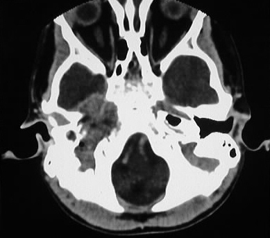
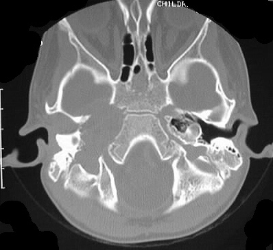
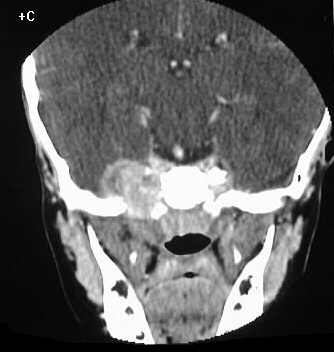
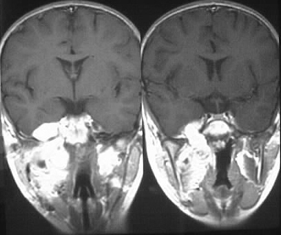
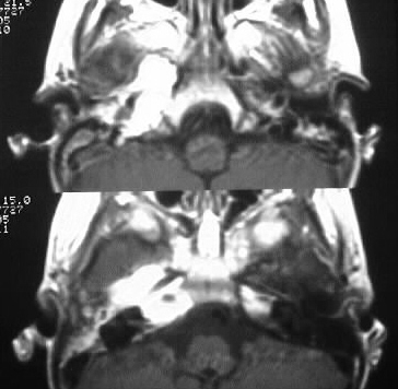
Findings:
Axial and coronal contrast enhanced CT demonstrates
a heterogenous strongly enhancing mass originating from the right masticator
space, with skull base invasion and intracranial extent. The skull base
foramina are destroyed in this region, but the tumor may have extended
through the foramen ovale initially. Coronal and axial enhanced T1 images
show the enhancing mass with infiltration of the right pterygoid muscles.
Differential Diagnosis:
Masticator space tumors include sarcomas (chondro-, rhabdo-,
osteo-), squamous cell carcinoma extension from oropharynx, lymphoma, schwannoma,
and metastasis.
Discussion:
Since the primary components of the masticator space
include muscles (mastication) , bone (mandible), and cartilage (TMJ), sarcomas
are relatively common in this location. Rhabdomyosarcoma is most commonly
seen in patients less than 10 years old, and has a propensity for skull
base invasion if it involves the nasopharynx or masticator space. The tumor
may be hemorrhagic. Response to chemotherapy and radiation may be favorable,
but the five year survival rate is less than 50% overall. Rhabdomyosarcomas
comprise approximately 10% of malignancies in patients less than 15 years
old.