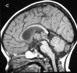
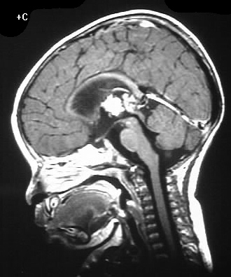
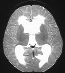

Choroid Plexus Papilloma
Findings:
A lobulated mass is present in the third ventricle which
is isointense to slightly hyperintense to gray matter on T1WI. This mass
enhances brightly. The lateral and third ventricles are dilated, without
obstructing lesion identified. There is evidence of transependymal flow.
Differential Diagnosis:
choroid plexus papilloma, exophytic glioma, less likely
intraventricular meningioma
Discussion:
-most common in first decade, 0.5% of all
intracranial neoplasms
-malignant degeneration 10-20% (imaging
nondiagnostic)
-trigone of lateral vent most common in
children
-4th vent most common in adult
-CSF seeding potential
-overproduction of CSF- hydrocephalus
-resorption also impaired due to hemorrhage,
increased protein content, +/- tumor pieces
imaging:
-iso/hyper on CT
-iso T1-marked enhancement