Adrenoleukodystrophy
Findings:
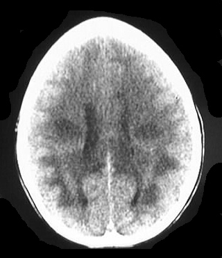
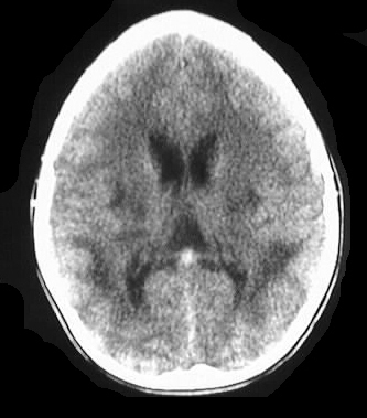
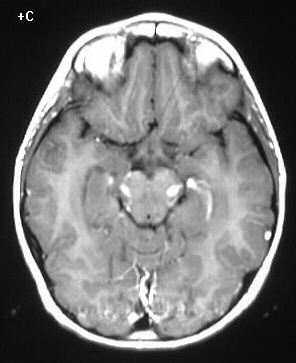
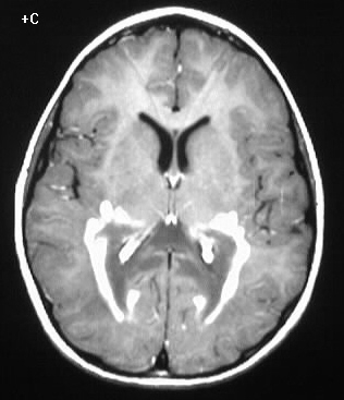
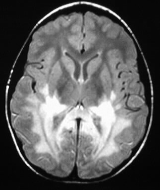
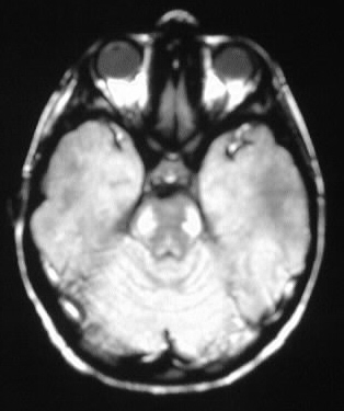
Axial CT images show patchy diffuse posterior white matter
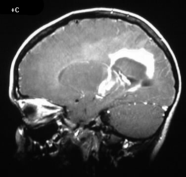
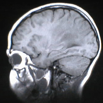
hypodensity. Multiple MR images show predominant signal abnormalities in
the parieto-occipital white matter and splenium of corpus callosum with
symmetric enhancement along the periphery. The signal abnormalities extend
into the brainstem.
Differential Diagnosis:
The signal characteristics are most consistent with ADL.
Globoid cell leukodystrophy may also have predominant abnormalities in
the parietal regions, but this characteristic enhancement pattern would
be unusual. MS and ADEM could rarely have this appearance, and would not
likely be this symmetric.
Discussion:
ADL represents a deficiency of lignoceroyl CoA ligase,
with accumulation of very long chain fatty acids in brain, adrenals, and
blood elements. It is inherited as x-linked recesive or autosomal recessive.
Pathologically, there is demyelination which advances in zones, with the
peripheral zone showing no inflammatory response. The intermediate zone
has active demyelination and inflammatory response, with the inner zone
showing necrosis, gliosis, and possible calcification. Numerous phenotypes
exist, the most common of which has clinical onset between 5-10 years,
with rapidly progressive neurologic decline and death. Imaging features
include symmetric posterior white matter involvement including the splenium,
with a rind of enhancement along the advancing edges. reference: Osborn, A; Tong, K. Handbook of Neuroradiology: Brain and
Skull. 1996: Mosby Year Book. pp. 521-523.