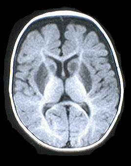
Findings:
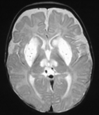
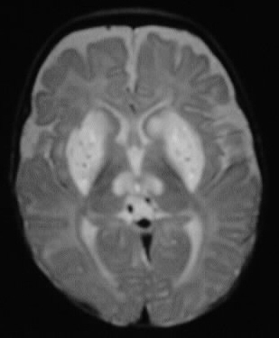
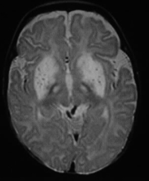
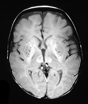
Multiple MR images show symmetric extensive abnormal signal in the putamen, globus pallidus, caudate, and thalamus. The signal characteristics are nonspecific T1 hypointensity and T2 hyperintensity. No mass effect is evident.
Differential Diagnosis:
The pattern fits best for Leigh disease. Other causes
of abnormal basal ganglia signal include carbon monoxide toxicity, methanol
intoxication, methylmalonic acidemia, cyanide intoxication, Wilson disease,
Hallervorden-Spatz. These other entities listed do not necessarily have
the same signal characteristics.
Discussion:
Leigh disease is a rare autosomal recessive mitochondrial
disorder that is also known as subacute necrotizing encephalomyelopathy.
The patients present clinically as infants with hypotonia, seizures, ophthalmoplegia,
and ataxia. The disease has a poor prognosis and causes symmetric necrosis
of the basal ganglia, periaqueductal gray, and brainstem. Mitochondrial
enzyme deficiencies include PDH complex, pyruvate carboxylase, and electron
transport chain.
reference: Osborn, A; Tong, K. Handbook of Neuroradiology: Brain and Skull 2nd edition.1996: Mosby-Year Book Inc. pp. 532.