Lyme Disease
Findings:
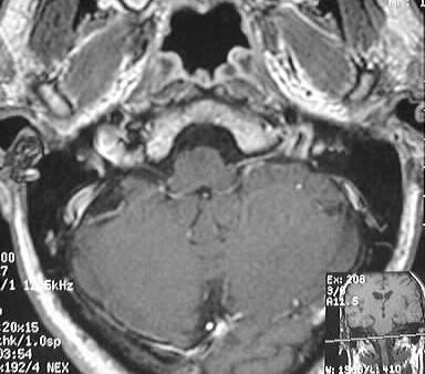
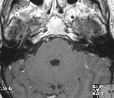
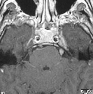
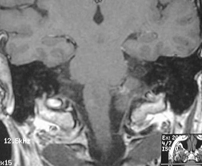
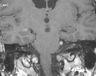
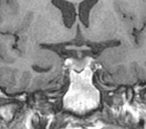
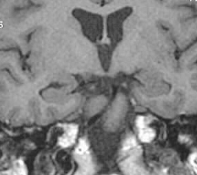
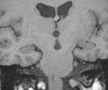
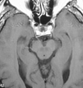
T1 postcontrast images show abnormal enhancement of multiple
cranial nerves.
Differential Diagnosis:
Lyme disease and sarcoidosis are the most common causes
of multiple cranial nerve enhancement. If the enhancement were limited
to a single unilateral cranial nerve, other etiologies such as schwannoma
or perineural tumor spread could be considered.
Discussion:
Lyme disease is caused by the spirochete Borrelia burgdorferi,
transmitted by the deer tick. Clinically the disease begins as a characteristic
rash centered on the tick bite known as erythema chronicum migrans. Systemic
dissemination is heralded by flulike illness and diffuse cutaneous lesions,
with the later development of neurologic, ocular and arthropathic symptoms.
The disease may also affect the heart with conduction abnormalities and/or
transient myopericarditis. Neurologic symptoms/signs develop in 10-15%
1-4 months after inoculation and include peripheral neuropathy, radiculopathy,
myelopathy, meningitis, movement disorders, and pain syndromes. Imaging
may simulate MS, with involvement of subcortical white mattter, brainstem,
and basal ganglia. Ring enhancement is occasionally seen, as is a cranial
neuritis or leptomeningitis. The imaging findings may be indistinguishable
from ADEM. Response to multiple antibiotic therapy is best if initiated
early in the course of the disease.