Glomus Jugulare
Findings:
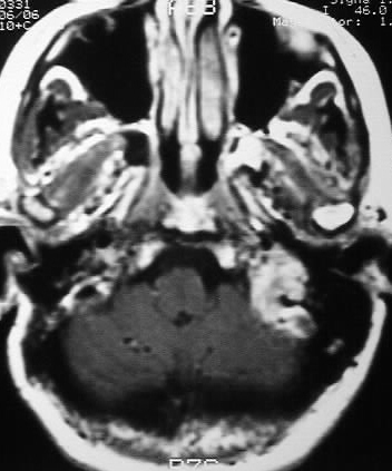
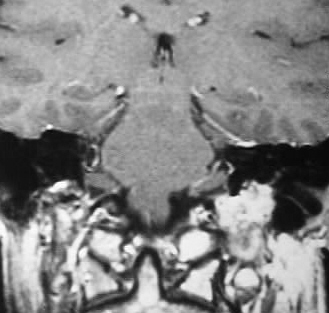
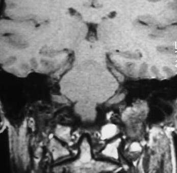
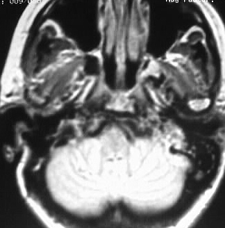
A strongly enhancing mass is present in the region of
the left jugular foramen, which exhibits heterogenous signal characteristics
and signal voids consistent with vascularity.
Differential Diagnosis:
Glomus jugulare, metastasis, schwannoma, neurofibroma,
meningioma.
Discussion:
Glomus Jugulare comprises approximately 90% of primary
tumors involving the jugular foramen. Flow voids are characteristic, with
a "salt and pepper" appearance and possible erosion of the jugular spine.
Attenuation and signal characteristics are nonspecific isodense CT/strong
enhancement CT+MR/hypo T1/hyper T2. The lesions are markedly hypervascular
on conventional angiography and preoperative embolization can be performed.
Arterial feeders are primarily ECA branches including posterior occipital,
ascending pharyngeal, and posterior meningeal. They resemble pheochromocytoma
histologically, but there is no association with NF or MEN, and endocrine
symptoms are uncommon. Recurrence is common, occuring in up to 50% of cases
(less for other glomus tumors).
reference: Osborn, A; Tong, K. Handbook of Neuroradiology: Brain and Skull 2nd ed. 1996: Mosby Year Book pp. 318-19.