Findings:
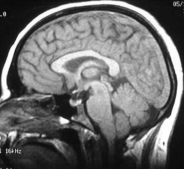
Sagittal T1 image shows absence of the normal flow void
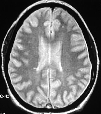
in the superior sagittal sinus. Axial PD/T2 weighted images show abnormal
flow signal in the transverse sinuses and signal abnormalities in the parafalcine
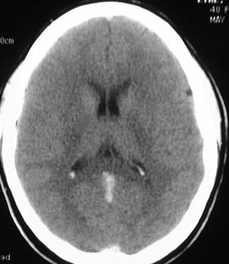
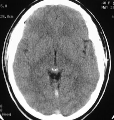
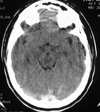
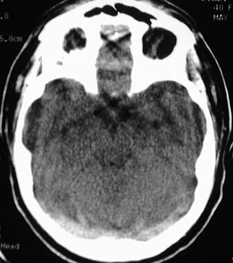
regions. Noncontrast CT demonstrates abnormal hyperdensity in the straight
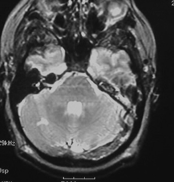
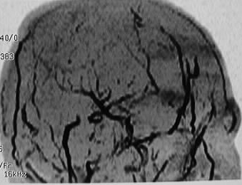
sinus, superior sagittal sinus, and transverse sinuses. MRV shows absence
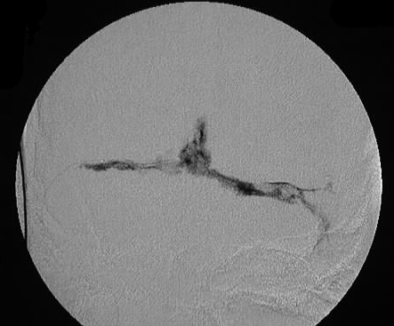
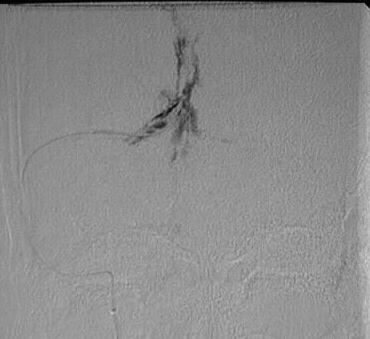
of flow signal in the dural sinuses. A catheter was positioned in the region
of the torcula, where contrast injection shows multiple filing defects
in the dural sinuses consistent with extensive thrombosis.
Discussion:
clinical:
-younger pts with headache, seizures, sudden focal
deficits
predisposing factors:
-hypercoagulable states, pregnancy, infection,
dehydration, meningitis, direct invasion from tumor
most common locations:
-transverse, superior sagittal, cavernous sinus
secondary signs:
-deep cortical or subcortical hemorrhage
-rounded configuration, may spare cortex
-not in specific arterial territory
pitfalls:
-CT: false (+) for empty delta sign if delay >
30 min.
-MR: slow flow may cause unusual signal combinations