
Straight sinus/Vein of galen thrombosis with bilateral thalamic infarction
Findings:
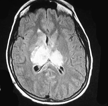
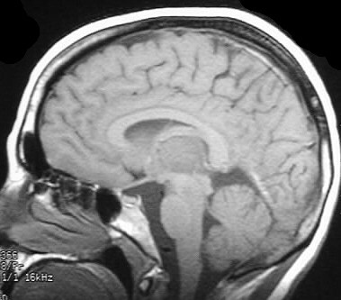
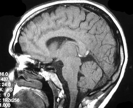
Axial T1 and T2 weighted images show extensive abnormal
signal with mass effect in the thalami, right internal capsule, and right
basal ganglia. The sagittal images show abnormal signal in the straight
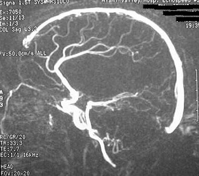
sinus/vein of galen region consistent with thrombosis. Lack of flow signal
in these areas is confirmed by phase contrast MIP image.
Differential diagnosis:
Bilateral signal abnormalities should raise the possibility
of dural sinus/deep venous thrombosis. Other etiologies of bilateral abnormal
thalamic signal abnormalities include "top of the basilar" syndrome (occlusion
of a large thalamoperforator that supplies both thalami), glioma, metastatic
disease, and less likely encephalitis.
Discussion:
Venous thrombosis may present with nonspecific symptoms
such as headache and seizures, or may be the cause of sudden focal deficits.
Predisposing factors include hypercoagulable states, pregnancy, infection,
dehydration, meningitis, and direct invasion from tumor. The transverse,
superior sagittal, and cavernous sinuses are the most common locations
of thrombosis. Careful attention should be paid to the density of the dural
sinuses on noncontrast head CT in the young adult population, with a low
threshold for further studies if there is a strong clinical suspicion of
thrombosis. MR venogram is the preferred definitive diagnostic test. Secondary
signs include deep cortical/subcortical hemorrhage and a rounded or flame
shaped configuration which may spare cortex. The signal abnormalities are
often not in a specific arterial territory.
Pitfalls:
-CT: false (+) for empty delta sign if delay >
30 min.
-MR: slow flow may cause unusual signal combinations