Trilateral retinoblastoma
Findings:
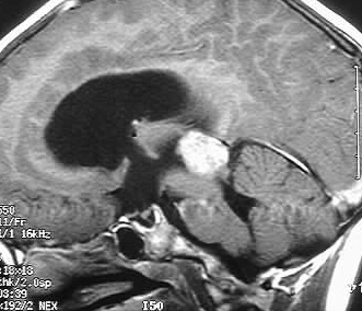
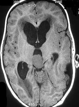
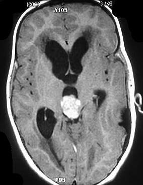
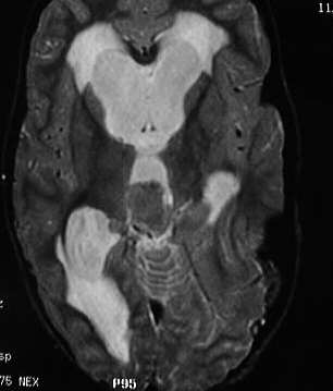
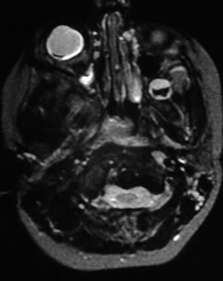
MR images show a strongly enhancing mass in the pinal
region which demonstrates isointensity on T2 weighted images. Moderate
hydrocephalus is present, associated with transependymal flow. The left
globe has been enucleated. A small focus of hypointensity is evident in
the right globe, which likely represents calcification.
Differential Diagnosis:
The differential diagnosis of a pineal region mass includes
germinoma, teratoma, and pineocytoma/blastoma. In this case, the associated
findings of previous retinoblastoma on the left and small calcified tumor
on the right would lead to a diagnosis of pineoblastoma.
Discussion:
Retinoblastoma is the most common primary intraocular
tumor of childhood, and should be a primary consideration in the differential
diagnosis of leukocoria. Intraocular calcifications in a child are indicative
of retinoblastoma until proven otherwise, and the other causes of
leukocoria (i.e. PHPV, Coat's dz, Toxocara, ROP) are rarely if ever calcified.
The tumors are thought to arise from primitive photoreceptor cells, and
25% are bilateral or multifocal. Invasion of the optic nerve portends a
poor prognosis. 10% are hereditary, associated with a deletion on chromosome
13q. These patients are at risk for developing pineal tumors, extracranial
sarcomas, and melanoma. When a patient with retinoblastoma develops a pineal
tumor, the term trilateral retinoblastoma is used.