Lhermitte-Duclos disease
Findings:
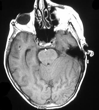
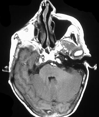
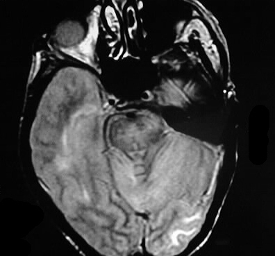
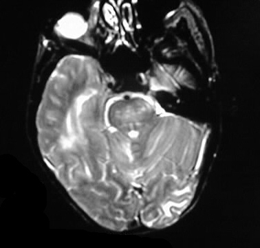
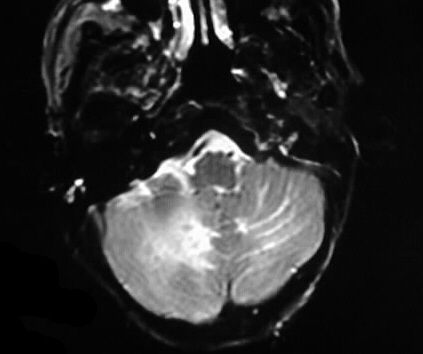
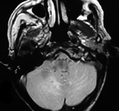
Multiple MR images show thickening of cerebellar folia
with striations and abnormal signal. No discrete enhancement is evident.
Differential diagnosis:
If no history was known, recent cerebellar infarction
could be considered. The appearance is characteristic for Lhermitte- Duclos
disease, and the distinction from infarction would not be a clinical dilemma.
Discussion:
Lhermitte-Duclos disease, also known as dysplastic gangliocytoma
of the cerebellum, presents as a focal or diffuse cerebellar hemisphere
mass with striation on MR. Symmetric thickening of the folia is a characteristic
feature. The lesions may represent a hamartomatous process rather than
a true neoplasm, but they do slowly enlarge. Histologically, there is absence
of purkinje cells with hyperplasia of molecular/granular layer and increased
myelin. Lhermitte-Duclos is also associated with Cowden Syndrome.