SLE Cerebritis
Findings:
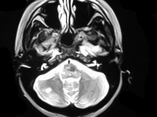
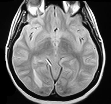
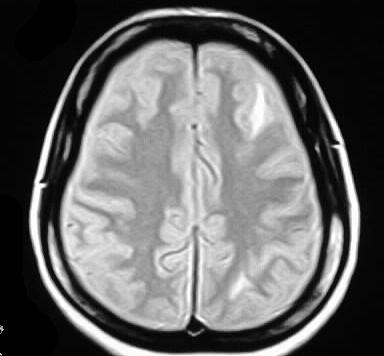
Scattered oval and flame shaped areas of abnormal signal
are present in the subcortical white matter bilaterally, involving the
left frontal lobe, bilateral occipital lobes, and right cerebellum. Abnormal
signal is evident in the brainstem as well.
Differential Diagnosis:
The appearance is relatively nonspecific and the differential
is broad. Multifocal ischemic foci are a possibility, due to vasculitis
or venous thrombosis. This would be an atypical distribution for MS or
ADEM. Metastatic disease would not likely have this configuration of lesions.
Discussion:
Up to 7% of patients with systemic lupus erythematosus
develop a CNS vasculitis. Pathologically, there is perivascular inflammation
and endothelial cell proliferation. Occipital involvement is characteristic,
with sparing of the periventricular regions. This helps to distinguish
the process from MS. Other manifestations of CNS lupus include atrophy,
infarcts, and hemorrhage. Infarcts in these patients may have several different
etiologies, including thrombotic (hypercoagulable state), embolic (vegetations),
or vasculitic. The vasculitic lesions occasionally respond to steroids.
reference: Grossman, R.I., Yousem, D.M. Neuroradiology: The Requisites. 1994:Mosby-Year Book. p. 115.