Mesial Temporal Sclerosis
Findings:
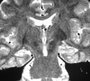
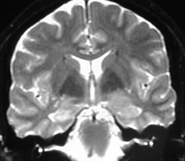
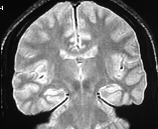
Coronal T2 weighted images show abnormal increased signal
in the hippocampus bilaterally, associated with white matter thinning in
these areas and decreased volume of the hippocampal formations. The changes
are slightly more pronounced on the right.
Discussion:
In the workup of chronic seizure disorder, attention
is focused to the medial temporal lobes. Mesial temporal sclerosis is the
most common process found, but other lesions seen include ganglioglioma,
cavernoma, dysembryoplastic neuroepithelial tumor, and astrocytoma. Diagnostic
criteria of MTS include volume loss and increased signal in the affected
temporal lobe. The process may be bilateral asymmetric or unilateral.