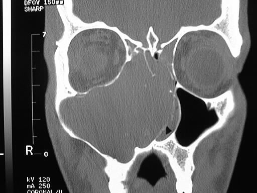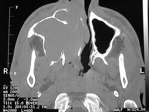

Findings:
Coronal and axial CT of the sinuses shows a large expansile
mass involving the right maxillary sinus with extension into the nasal
cavity and ethmoids.
Differential Diagnosis:
The appearance is somewhat nonspecific. Since the lesion
expands and remodels bone rather than destroys it, malignancies such as
squamous cell carcinoma, esthesioneuroblastoma, and salivary gland tumors
would be relatively unlikely. Reasonable possibilities include inverting
papilloma, antrochoanal polyp, or mucocele.
Discussion:
Inverting papillomas are most common in middle aged males
and present with unilateral nasal obstruction. The tumors are histologically
benign, but foci of squamous cell carcinoma within the lesion are not uncommon.
The tumors usually arise from the lateral nasal wall and extend into the
maxillary and ethmoid sinuses. Bone destruction may be seen and is usually
the result of pressure necrosis rather than tumor permeation. The lesions
enhance homogenously, and may have intermediate/low signal on T2WI. The
maxillary sinus infundibulum is typically markedly widened. The etiology
of the lesions is unknown.