Cysticercosis
Findings:
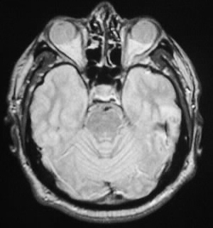
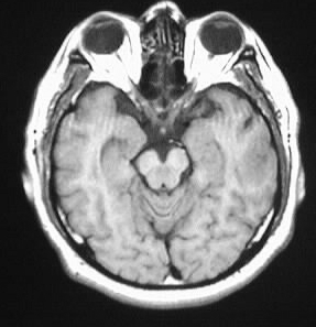
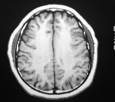
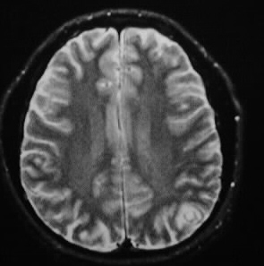
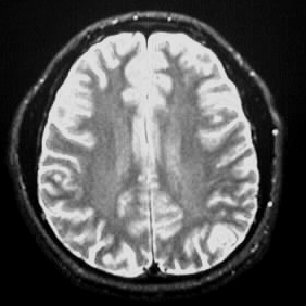
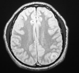
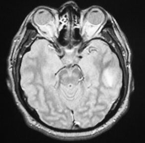
Multiple MR images show scattered subcortical signal
abnormalities in the left temporal lobe and left parietal region. These
lesions have hypointense rims, near fluid signal centers, and surrounding
edema.
Differential Diagnosis:
cysticercosis, mets, abscesses
Discussion:
Cysticercosis is the most common worldwide CNS infection.
CNS lesions are present in 60-90% of infected individuals. The disease
is contracted by ingesting the ova of the pork tapeworm Taenia solium in
contaminated water. Clinically, seizures are the most common presentation.
In endemic areas, seizures in a young adult are due to cysticercosis until
proven otherwise. The disease is treated medically with praziquantal or
albendazole, with occasional surgery for shunting or removal of intraventricular
lesions.
Stages:
-vesicular: cyst with nodule, no edema,
live larva
-colloid vesicular: dying larva, ring
enhancement with edema
-granular nodular- healing, calcified
enhancing cyst
-nodular calcified- small calcified
nodule without edema or enhancement
Imaging:
-lesions may be parenchymal, intraventricular,
meningeal, or racemose (basilar cisterns)
-multiple low density CT, CSF iso on MR,
peripheral gray/white junction
-acute encephalitis with rupture
-ring enhancing with edema, increased density/signal,
eventually calcifies
-intraventricular lesions may acutely obtruct
-racemose form with grapelike clusters in
basal cisterns-->hydrocephalus
reference: Osborn, A. Tong, K. Handbook of Neuroradiology: Brain and
Skull. 1996:Mosby-Year Book. pp. 490-92.